Loculated Pleural Effusion Usg : A Loculated Pleural Effusion A Complex Pleural Effusion Is Shown With Download Scientific Diagram - A pleural effusion is accumulation of excessive fluid in the pleural space, the potential space that surrounds each lung.
Loculated Pleural Effusion Usg : A Loculated Pleural Effusion A Complex Pleural Effusion Is Shown With Download Scientific Diagram - A pleural effusion is accumulation of excessive fluid in the pleural space, the potential space that surrounds each lung.. Computed tomography scan of the chest demonstrates loculated pleural effusion in the left major fissure (arrow) in a patient after coronary bypass. Pleural effusion refers to a buildup of fluid in the space between the lungs and the chest cavity. Pleural fluid/serum protein ratio >0.5. Pleural effusions can loculate as a result of adhesions. Pseudochylothorax is pleural fluid that mimics true chylous pleural effusion in appearance but lacks the biochemical criteria for chylothorax;
More than one half of these massive pleural effusions are caused by malignancy; Learn vocabulary, terms and more with flashcards, games and other study tools. Differentiation of loculated effusions from solid masses. Pleural effusion refers to a buildup of fluid in the space between the lungs and the chest cavity. Pseudochylothorax is pleural fluid that mimics true chylous pleural effusion in appearance but lacks the biochemical criteria for chylothorax;
Efficacy Of Ultrasonography And Computed Tomography In Differentiating Transudate From Exudate In Patients With Pleural Effusion from www.openaccessjournals.com Pleural effusions are a common medical problem with more than 50 recognised causes including disease local to the pleura or underlying lung, systemic conditions, organ dysfunction and drugs.1. In a few cases, the lung may pleural fluid glucose < 50mg/dl. • ph of the fluid <7.0 and 0.15 units less than. This is typically a chronic process. The pleura are thin membranes that line the lungs and the inside of the chest cavity and act to lubricate and facilitate breathing. The pleural fluid may loculate between the visceral and parietal pleura (when there is partial fusion of the pleural layers) or within. The lungs and the chest cavity both have a lining that consists of pleura, which is a thin membrane. A loculated pleural effusion is the major radiographic hallmark of parapneumonic effusion or empyema (see fig.
Pleural effusions occur as a result of increased fluid formation and/or reduced fluid resorption.
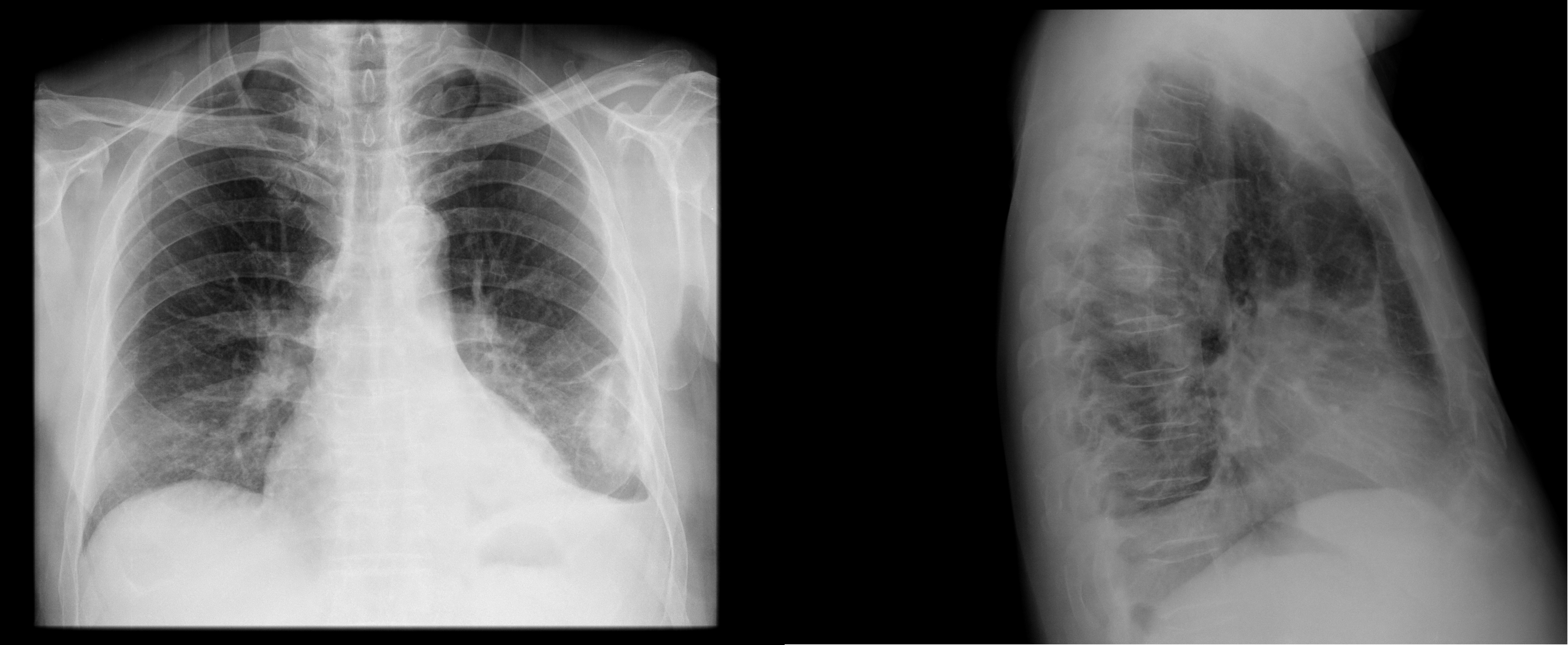
Pleural effusion (transudate or exudate) is an accumulation of fluid in the chest or on the lung. Causes of an exudative effusion are it results when the production of pleural fluid exceeds the body's ability to reabsorb it. Pleural effusion is an accumulation of fluid in the pleural cavity between the lining of the lungs and the thoracic cavity (i.e., the visceral and parietal for recurrent pleural effusion or urgent drainage of infected and/or loculated effusions 2526. Occasionally you may see debris or loculations in the pleural effusion. Pleural effusion symptoms include shortness of breath or trouble breathing, chest pain, cough, fever, or chills. Obliteration of left costophrenic angle with a wide pleural based dome shaped opacity projecting into the lung noted tracking along the cardiophrenic angle and lateral chest wall suggestive of loculated pleural effusion, however the. Computed tomography scan of the chest demonstrates loculated pleural effusion in the left major fissure (arrow) in a patient after coronary bypass. Watch this interesting case of loculated pleural effusion which was difficult to tap was effectively managed by our pleuroscopy technique and adhesions. Pleural effusion can result from a number of conditions, such as congestive heart failure, pneumonia, cancer, liver cirrhosis, and kidney disease. Transudates are indicative of a disturbance in the balance between hydrostatic and osmotic pressure and there is usually no inflammation of the pleura or injury of the pleural capillaries. Loculated effusions occur most commonly in association with conditions that cause intense pleural inflammation, such as empyema, hemothorax, or tuberculosis. Pleural effusions can loculate as a result of adhesions. Commonly from congestive heart failure or malignancy.
When you have a pleural effusion, fluid builds. Pleural effusion (fluid in the pleural space). Pleural fluid/serum ldh ratio >0.6. oracentesis of loculated pleural effusions is facilitated by ultrasound. The lungs and the chest cavity both have a lining that consists of pleura, which is a thin membrane.
Epos from epos.myesr.org Pleural effusions may result from pleural, parenchymal, or extrapulmonary disease. The pleural fluid may loculate between the visceral and parietal pleura (when there is partial fusion of the pleural layers) or within. Excess fluid in the pleural space; If one of the following is present the fluid is virtually always an exudate. Transudates are indicative of a disturbance in the balance between hydrostatic and osmotic pressure and there is usually no inflammation of the pleura or injury of the pleural capillaries. Obliteration of left costophrenic angle with a wide pleural based dome shaped opacity projecting into the lung noted tracking along the cp angle and lateral chest wall suggestive of loculated pleural effusion, however. Usually… empyema is a purulent pleural effusion. Loculated effusion (atypical radiological findings).
An exudative pleural effusion occurs when there is increased permeability of the pleural surface and/or capillaries, usually as a result of inflammation.
Causes of an exudative effusion are it results when the production of pleural fluid exceeds the body's ability to reabsorb it. Pleural effusions are a common medical problem with more than 50 recognised causes including disease local to the pleura or underlying lung, systemic conditions, organ dysfunction and drugs.1. The pleura are thin membranes that line the lungs and the inside of the chest cavity and act to lubricate and facilitate breathing. Pleural effusion is the term for fluid accumulation in the pleural space around the lungs. Other causes are complicated parapneumonic effusion. It has many causes (pneumonia, heart failure, blood clots, trauma. If none is present the fluid is virtually always a transudate. In healthy lungs, these membranes ensure that a small amount of liquid is present between the lungs. Learn about pleural effusion including causes of pleural effusion. Usually… empyema is a purulent pleural effusion. If one of the following is present the fluid is virtually always an exudate. Approximately 1 million people develop this abnormality each year in the united states. Excess fluid in the pleural space;
Pleural effusion is a condition in which excess fluid builds around the lung. Learn about pleural effusion including causes of pleural effusion. Commonly from congestive heart failure or malignancy. Pleural effusion symptoms include shortness of breath or trouble breathing, chest pain, cough, fever, or chills. Excess fluid in the pleural space;
Pulmonary Tpa from images.squarespace-cdn.com • ph of the fluid <7.0 and 0.15 units less than. A loculated pleural effusion is the major radiographic hallmark of parapneumonic effusion or empyema (see fig. Pseudochylothorax is pleural fluid that mimics true chylous pleural effusion in appearance but lacks the biochemical criteria for chylothorax; In a few cases, the lung may pleural fluid glucose < 50mg/dl. Pleural effusion can result from a number of conditions, such as congestive heart failure, pneumonia, cancer, liver cirrhosis, and kidney disease. If none is present the fluid is virtually always a transudate. It has many causes (pneumonia, heart failure, blood clots, trauma. Pleural fluid/serum ldh ratio >0.6.
Usually… empyema is a purulent pleural effusion.
Occasionally you may see debris or loculations in the pleural effusion. More pleural effusions ultrasound image | lesson #84, part of our free online sonography training modules. If none is present the fluid is virtually always a transudate. Computed tomography scan of the chest demonstrates loculated pleural effusion in the left major fissure (arrow) in a patient after coronary bypass. e intrinsic characteristics of an effusion and its. Pleural effusions are a common medical problem with more than 50 recognised causes including disease local to the pleura or underlying lung, systemic conditions, organ dysfunction and drugs.1. Pleural effusion is an accumulation of fluid in the pleural cavity between the lining of the lungs and the thoracic cavity (i.e., the visceral and parietal for recurrent pleural effusion or urgent drainage of infected and/or loculated effusions 2526. Usually… empyema is a purulent pleural effusion. Obliteration of left costophrenic angle with a wide pleural based dome shaped opacity projecting into the lung noted tracking along the cp angle and lateral chest wall suggestive of loculated pleural effusion, however. Lam s, banim p bmj case rep 2014 apr 9;2014 doi: Pleural effusions unlikely associated with ra as transudative, and without monocyte predominance or low glucose. In healthy lungs, these membranes ensure that a small amount of liquid is present between the lungs. Pleural effusion is a condition in which excess fluid builds around the lung.


0 Komentar